
Identification
Wood & Charcoal Identification in Southern Maryland
By Harry Alden
Wood & Charcoal Anatomy Basics
|
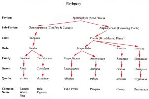 |
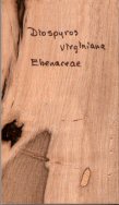
| The phylogeny of 5 woods: Eastern White Pine (Pinus strobus), Bald Cypress (Taxodium distichum), Tulip Poplar (Liriodendron tulipifer), Pawpaw (Asimina triloba), Cherry (Prunus serotina) and Persimmon (Diospyros virginiana). |
Gross Structural Features
Planes of Section
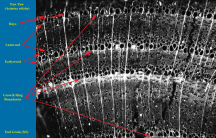
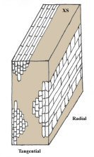
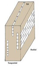
Wood is a three-dimensional material, with the cells of tissues oriented in three different planes of view (charcoal) or planes of section (wood). The plane that cuts across the trunk or branch perpendicular to the grain is called the transverse or cross section (XS). The plane that runs longitudinally from the center of the trunk or branch to the bark is called the radial section (R). The plane that is perpendicular to the radial plane and is tangential to the growth rings is called the tangential section (T).
Click
image to view larger image.
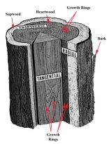 |
| Diagram showing planes of section, bark, growth rings, sapwood and heartwood. |
Growth Increments (Rings)
For all ring porous and semi-ring porous hardwoods and some softwoods in Calvert County, there is enough of a difference between the earlywood of one year and the previous latewood to produce annual growth rings visible to the naked eye. For the most part, the age of a tree can be calculated by counting the growth rings either from a stump or from other cross sections of the trunk. The inner part of the growth ring formed first in the growing season is called earlywood and the outer part formed later in the growing season, latewood. Earlywood is characterized by cells having relatively large cavities and thin walls, while latewood cells have smaller cavities and thicker walls. The transition from earlywood to latewood may be noticeable or gradual, depending on the taxon and the growing conditions when it was being formed.
Click on each image to view larger image.
 |
 |
 |
||
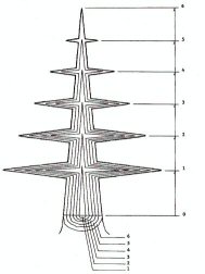
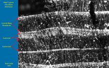
| Schematic diagram of a tree stem cut away to the center, exposing 6 growth sheaths or growth rings (bottom). The top of each sheath shows the height of the tree at the end of each growing season (right). From: Foulger, A.N. 1969. Classroom Demonstrations of Wood Properties. USDA/FPL. (Public Domain) | End grain of Bald Cypress (Taxodium distichum) charcoal showing several growth rings. | End grain of Pawpaw (Asimina triloba) charcoal showing several growth rings. |
Earlywood & Latewood
During the course of a growing season, the tree will produce cells early in the season (earlywood) and cells towards the end of the season (latewood). During stress (drought, defoliation, fire) some trees produce so little earlywood that the growth ring disappears into the previous year’s latewood.
Click
here to view larger image.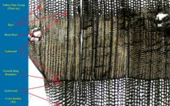 |
Click
here to view larger image.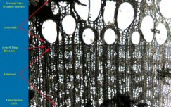 |
|
| Cross-section
of Yellow Pine Group (Pinus sp.), showing 2 partial growth rings |
Cross-section
of Trumpet Vine (Campsis
radicans),showing the growth ring boundary, the earlywood (top) and the latewood (bottom) of the previous growth ring. |
Sapwood and Heartwood
Sapwood is located between the bark and the heartwood. The sapwood contains both living and dead cells and functions primarily in the storage of food and the mechanical transport of water or sap. The sapwood layer may vary in thickness and in the number of growth rings contained in it. Sapwood commonly ranges from 1-1/2 to 2 inches in radial thickness. As a rule, the more vigorously growing trees have wider sapwood layers. Many second growth trees of merchantable size consist mostly of sapwood.
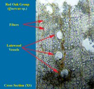
In general, heartwood is comprised of inactive cells that neither conduct water or store food. Frequently these extractives darken the heartwood and give species such as black walnut and cherry their characteristic color. Heartwood extractives in some species such as black locust, western redcedar, and redwood make the wood resistant to fungi or insect attack. Sapwood of all species, however, is not resistant to decay. In some species, such as the ashes, hickories, and certain oaks, the pores (vessels become plugged to a greater or lesser degree with ingrowths known as tyloses. Heartwood having pores tightly plugged by tyloses, as in white oak, is suitable for tight cooperage, since this prevents the passage of liquid through the pores. Tyloses also make impregnation with liquid preservatives difficult.
Click image
to view larger image.
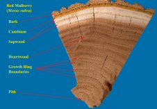 |
| End grain view of Red Mulberry (Morus rubra), showing bark. |
    |
|
Anatomical Features
Wood Cells
Wood cells make up the structural elements of wood and are composed of various sizes and shapes. Dry wood cells may be empty or part filled with deposits, such as gums and resins. Vessels can be occluded with tyloses, neighboring parenchyma cells that balloon into the vessel, closing it up. The main wood cell types that function in transport and support (are elongated and pointed at the ends) are the fibers and tracheids. In addition to fibers, hardwoods have cells of relatively large diameter that function in the transport of fluids, the vessels (or pores). The transport function is also performed by the tracheids in softwoods and hardwoods. Both hardwoods and softwoods have cells that are grouped into tissues oriented horizontally in the radial direction from the pith towards the bark. These cells conduct fluids radially and are called rays. The rays are most easily seen on end-grain (XS) or quartersawn (R) surfaces. Wood also has other cells, known as longitudinal or axial parenchyma that function mainly for the storage of food, extractives and/or crystals. All of these cell types and their arrangement into tissues, three dimensionally, constitutes the basis for the microscopical description and identification of woods.
Cells Involved (Softwoods vs. Hardwoods
Softwoods
Click here to view larger image.
 |
| Diagram showing types of softwood cells and the cambial initials that produced them. The cambial initials are shown from approximately the tangential aspect. The two types of pit arrangement are shown in the tracheids. Modified from Jane, F.W. 1970. The Structure of Wood. |
Axial/Vertical Tracheids
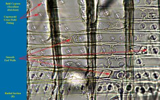
Axial tracheids are the main cell type in softwoods, comprising over 90% of cells in the wood. Their radial diameter changes depending on growth rates, the order formed during the growing season (earlywood tracheids are larger than latewood tracheids) and environmental conditions. In some softwoods, like Bald Cypress (Taxodium distichum) and Sequoia (Sequoia sempervirens), the first formed tracheids are always wide enough to develop inter-tracheid pitting that has more than one row of pits and is of diagnostic value.
| Radial section of Bald Cypress (Taxodium distichum) showing biseriate (2-wide) bordered pits (arrows) in the axial tracheids. |
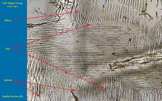
Spirals
A few softwoods have spiral thickenings on the walls of their tracheids and these can be diagnostically significant. However, archaeological or degraded wood frequently has spiral checks in the cell walls, appearing like spiral thickenings.
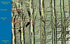
Ray Tracheids
Ray tracheids are like small axial tracheids oriented radially. Like axial tracheids, they have circular bordered pits (CBP), just smaller. The rays tracheids can have smooth walls like White Pine Group or the walls have pointed projections (dentate) within them as in the Yellow and Red Pine Groups. The main difference between spruce and larch is the shape of the cross section CBP in the ray tracheids.
Click each image to view larger image.
 |
 |
|
| Radial section of White Pine Group (Pinus sp.) showing 1 row of ray tracheids and 5 rows of ray parenchyma cells below. | Radial section of Yellow Pine
Group Pinus sp.) showing 3 rows of ray tracheids. |
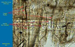
Axial/Vertical Parenchyma
Axial parenchyma in softwoods is usually present diffusely in the latewood part of the growth ring. They usually contain colored contents and are easy to see at low magnifications. In tangential view, the walls separating adjacent cells will either be smooth or nodular.
Click here to view larger image.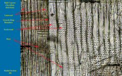 |
| Tangential section of Bald Cypress (Taxodium sp.) showing axial parenchyma with nodular endwalls. |
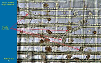
Ray Parenchyma
Ray parenchyma in softwoods have either smooth end walls or end walls with distinct nodules on them (nodular end walls).
Click each image to view larger image.
 |
 |
|
| Radial section
of Hemlock (Tsuga spp.) showing ray parenchyma cells with nodular end walls. |
Radial section of Bald Cypress (Taxodium distichum) showing ray parenchyma cells with smooth end walls. |
Epithelial Cells of Resin Canals
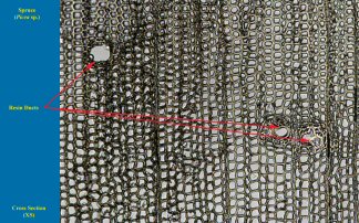
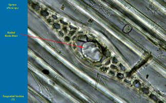
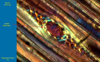
In many softwood and some hardwoods, cells are arranged axially or radially into canals or ducts which produce resins, tannins and other secondary compounds used in protection against pathogens like insects and fungi. In softwoods, the resin canals are easily seen in both Xs and T views. The can have thick cell walls (epithelia) as in Spruce (Picea sp.) and Larch (Larix sp.) or thin walls as in Pines (Pinus spp.).
Click each image to view larger image.
 |
 |
|
| Cross-section
of Pine (Pinus sp.) showing a single resin duct with thin-walled epithelial cells. |
Cross-section of
Spruce (Picea sp.) showing 3 resin ducts with thick-walled epithelial cells. |
 |
 |
|
| Tangential section of Pine (Pinus sp.) showing a single resin duct with thin-walled epithelial cells. Lucky hand section that retained epithelia. | Tangential
section of Pine (Pinus sp.) showing a single resin duct with thin-walled epithelial cells. Normal hand sections with torn out epithelia. |
| |
|
|
| Radial
section of Hemlock (Tsuga spp.) showing ray parenchyma cells with nodular end walls. |
Radial section of Bald Cypress (Taxodium distichum) showing ray parenchyma cells with smooth end walls. |
 |
 |
|
Tangential section of
Spruce (Picea sp.) |
Tangential
section of Spruce (Picea sp.) showing a single resin duct with thick-walled epithelial cells. (Polarized Light). |
Cross-Field Pits (not cells)
When rays intersect with axial tracheids, the cells that are adjacent to each other have intercellular connections called cross-field pits. They are where the axial system and ray system cross and are important diagnostic characters.
Click on image to view larger image.
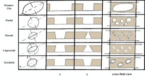 |
| Diagrammatic representations of pitting on radial walls of ray parenchyma cells in softwoods. [x - pits in section cut along the x axis; y - pits in section cut along the y axis; cross-field view - pits as seen in surface view in a cross-field] Modified from Jane, F.W. 1970. |
The Structure of Wood
Hardwoods
Cell Types
Hardwoods are distinguished form softwoods by having (usually) vessels and fibers in addition to parenchyma and tracheids. A few, primitive hardwoods are vessel-less. The more primitive tracheid is a good conductor of fluids and is good as a support element. Fibers evolved to be a poor conductors, but an excellent supportive elements, while vessels evolved for excellent conduction but poor support. Parenchyma can contain colored compounds or crystals or be segmented and aligned horizontally in a tissue. Vessel elements are connected vertically (along the grain) into vessels, that have perforation plates of various types. All four types of cells have evolved into different sizes, shapes and ornamentations in separate taxa and are the basis of all accurate microscopic identifications.
Click on image to view larger image.
 |
|
 |
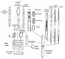
| Diagram showing types of hardwood cells and the cambial initials that produced them. The several types of perforations are shown in the vessels. Modified from Jane, F.W. 1970. The Structure of Wood. | The evolution of fibers and vessel members from tracheids (division of labor/function). |
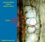
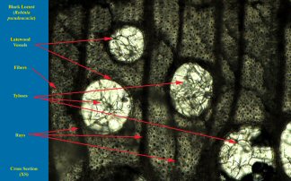
Tyloses
In some hardwoods, during heartwood formation, vasicentric parenchyma balloon into the interior of adjacent vessels elements. The tyloses efficiently block the unused vessels as passage ways for pathogens like insects and fungi. The presence of tyloses in white oak allows it to be used as “tight cooperage” that will not leak fluids (wine, whiskey, beer, water, etc.), as opposed to red oak that is used as “slack cooperage” for dry goods. Tyloses can appear small or large, with thick or thin walls and is a good diagnostic character when used properly.
Click on image to view larger image.
|
|
 |
|
| Radial
section of American Black Walnut (Juglans nigra) showing tyloses. |
Radial
section of Black Locust (Robinia pseudoacacia) showing tyloses. |
Perforation Plates
Vessels are distinguished from tracheids by having perforations at their ends to allow for efficient transport of fluids. These openings can be completely open (simple perforation) or can have bars (ladder rungs) across it (scalariform perforation). The bars can be few and thick or many and thin and are of diagnostic value.
Click on image to view larger image.
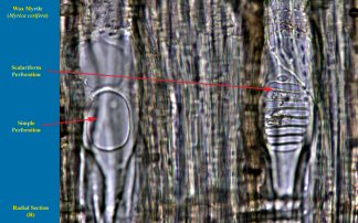 |
| Radial section of Wax Myrtle (Myrica cerifera) showing 2 vessels, 1 with a simple perforation and another with a scalariform perforation. |
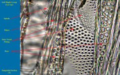
Inter-vessel (IV) Pits
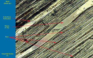
The connections between adjacent vessel members are called inter-vessel (IV) pits. They are easily seen in tangential view, except when the vessel distribution is solitary. They vary in size from the limits of most optical microscopes (2µ) to linear pitting that nearly spans the vessel (to 50µ).
| PIT SIZE | EXAMPLE |
| Minute (2 – 4µ) | Birch |
| Small (5-7µ) | Mountain Laurel |
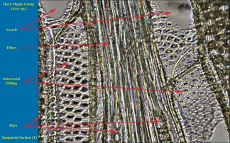
| Medium (8 – 10µ) | Maple |
| Large (> 10µ), includes linear | Walnut |
Click on each image to view larger image.
 |
 |
|
| Tangential
section of Birch (Betula sp.) showing minute inter-vessel pits (2 – 4µ). |
Tangential
section of Maple (Acer sp.) showing inter-vessel pits of medium size (5 – 10µ). |
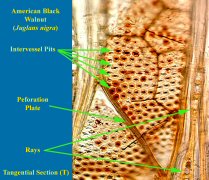 |
| Tangential section of Walnut (Juglans
sp.) showing large inter-vessel pits (arrowheads) of 10-16µ. |
IV. Arrangement
The arrangement of IV pits can be opposite each other, alternating horizontally or composed of long openings, spanning the vessel member (linear or scalariform).
| Type | Example |
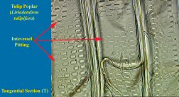
| Opposite | Tulip Poplar |
| Alternate | Maple |
| Linear/Scalariform | Sweet Gum |
Click on
each image to view larger image.
 |
 |
|
| Tangential section of Tulip Poplar
(Liriodendron tulipifera) showing opposite inter-vessel pits (arrows). |
Tangential section of Maple (Acer sp.) showing alternate inter-vessel pits arrows). |
 |
| Tangential section of Red Gum (Liquidambar styraciflua) showing linear inter-vessel pits arrowheads). |
Ray-Vessel (RV) Pitting
The connections between vessel elements and adjacent ray parenchyma cells are called ray-vessel (RV) pits. It can be the same as the IV pitting or much larger (salicoid) to linear.
Click on each image to view larger image.
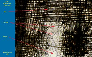 |
| Radial section of Elm (Ulmus
sp.) showing ray-vessel pits. |
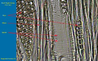
Spirals
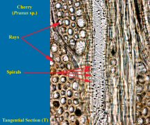
Some hardwoods have spiral thickenings on the walls of their vessel members. A few taxa have spirals that are limited to vessel element tips. Some spirals can be thin and evenly arranged, not passing through inter-vessel (IV) pitting, like Maple (Acer sp.). Other spirals are thicker and coarsely laid down and do pass through IV pitting like Cherry (Prunus sp.), Basswood (Tilia sp.) and Holly (Ilex sp.). However, archaeological or degraded wood frequently has spiral checks in the cell walls, appearing like spiral thickenings.
Click on each image to view larger image.
| Fine Spirals (Maple) | Coarse Spirals (Cherry & Basswood) | |
 |
 |
|
| Tangential section of Maple (Acer sp.) showing fine spirals. |
Tangential sections of Cherry (Prunus serotina) showing coarse spirals. |
Tracheids
Although softwoods are composed mostly of tracheids, hardwoods can have both vascular tracheids (like softwoods) and vasicentric tracheids that surround vessels in woods like Oak.
Click on image to view larger image.
 |
| Tangential section of Oak (Quercus sp.) showing vasicentric tracheids (arrowhead) with circular bordered pits. |
Fibers
The main type of supporting cell in hardwoods is the libriform fiber. These cells can have partitions within them (septate) or not (non-septate). There is also a group of cells (fiber tracheids) that are intermediate in structure between fibers and tracheids. These cells can also be categorized as septate or non-septate.
Click on each image to view larger image.
 |
 |
|
| Cross section of Oak Group
(Quercus sp.), showing moderately thick walled fibers. |
Cross section of Black Locust (Robinia pseudoacacia) showing extremely thick walled fibers. |
Axial Parenchyma
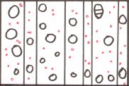
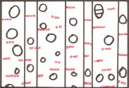
Parenchyma strands that are oriented vertically, or with the grain, within the wood are called axial parenchyma. They are grouped into two broad categories; apotracheal parenchyma and paratracheal parenchyma. Apotracheal means “without vessels” and is axial parenchyma that does not touch any vessels in cross section. Types of apotracheal parenchyma include diffuse, reticulate or broken banded and banded. Paratracheal means “around the vessels” and is axial parenchyma that either completely or partially surrounds vessels in cross section. Types of paratracheal parenchyma include vasicentric (“surrounding vessel”), aliform (winged) and confluent (coalesced wings).
Click on each
image to view larger image.
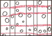
Diffuse Reticulate
Banded
Diagram of different types of apotracheal
parenchyma in hardwoods, as seen in cross-section.
Vessels are represented by circles, rays by vertical lines and
parenchyma by red areas.
Click on each
image to view larger image.
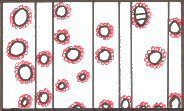
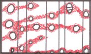
Vasicentric Aliform
Confluent
Diagram of different types of paratracheal
parenchyma in hardwoods, as seen in cross-section.
Vessels are represented by circles, rays by vertical lines and
parenchyma by red areas.
Rays
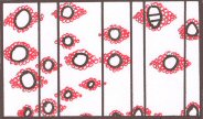
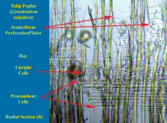
Rays are composed of parenchyma cells in hardwoods and of parenchyma and ray tracheids in softwoods. In hardwoods, rays are categorized by cell width and heterogeneity. Rays can be one cell wide in tangential view (uniseriate) or many cells wide (multiseriate). They can be of just one type of cell (square or procumbent), called homocellular, or it can have both procumbent cells in the body of the ray and one to many rows of squares or upright cells along the periphery (heterocellular).
Click on each
image to view larger image.
Uniseriate Rays Multiseriate Rays
Homocellular Rays Heterocellular Rays
Different types of rays in hardwoods as seen in tangential, radial
and cross sections.
Click on each image to view larger image.
 |
 |
|
| Radial section of Maple (Acer sp.)
showing a homocellular ray composed exclusively of procumbent cells. |
Radial section of
Tulip Poplar Liriodenron tulipifera) showing a heterocellular ray composed of procumbent cells (top half of figure) and uprights (arrowheads). |
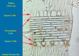 |
| Radial
section of Willow (Salix spp.) showing a heterocellular ray composed of procumbent cells (bracket) and uprights (arrowheads). |
|
|
Thank you for visiting our website. If you have any questions, comments, or new information to share, please contact us at [email protected]. Copyright © 2009 by |
|